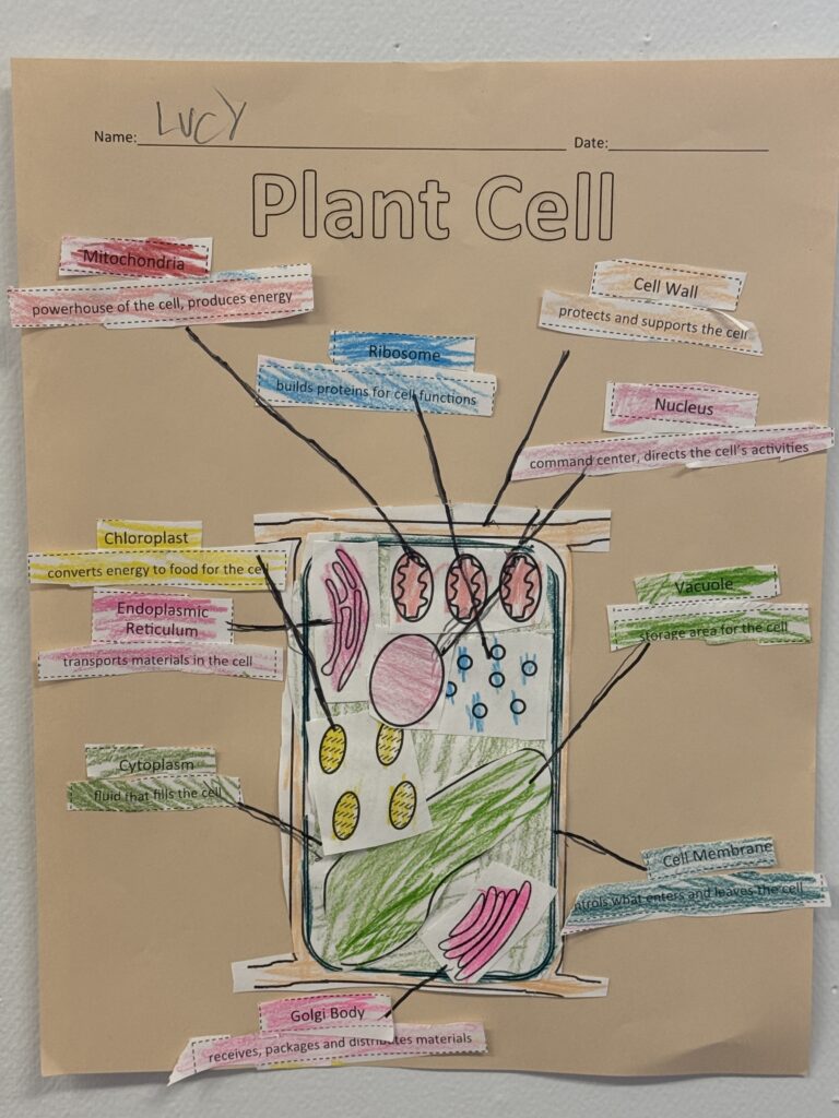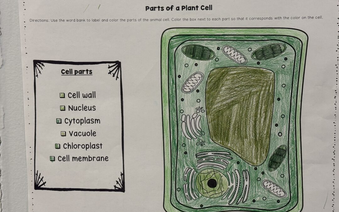
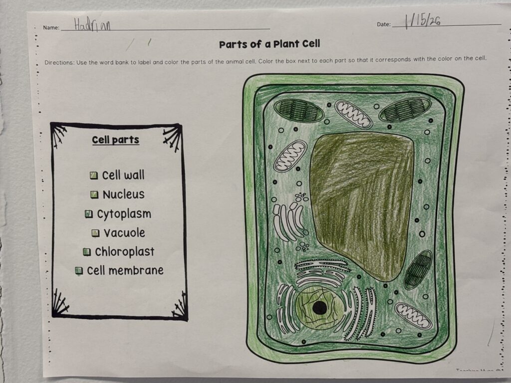
This winter and spring of 2026, students in grades 6th, 7th and 8th have been learning about plant and animal cells. We started our cellular exploration by looking at many parts of the cell and learning about the most important parts that help the cell function. Students identified and labeled parts of the plant cell including: ribosomes, nucleus, cell membrane, central vacuole, mitochondria, chloroplasts, endoplasmic reticulum, Golgi body, cytoplasm and cell wall.
We then took a microscopic look at a piece of an onion. Students practice making wet mount slides and using the different powers of the microscope to see the onion on the cellular level. As we moved on to animal cells, students were able to compare and contrast the plant cell to the animal cell. One of the biggest differences students noticed was there was no cell wall in an animal cell. To see animal cells under the microscope, we scraped cheek cells from our mouths. Each student made their own wet mount slide and also dyed the cells blue with methylene blue to help observe under the microscope. Students also got a chance to look at a piece of our gecko, Juliana’s, shredded skin. We are now observing all different types of items under the microscopes and comparing them to the cells we have seen.